When two is better than one… Double-layered amniotic membranes improved OSD and visual acuity.
When the ocular surface refuses to cooperate, clinicians tend to reach for every tool in the box. A recent study suggests one familiar option, used a little differently, may deserve renewed attention.
A double-layered dehydrated amniotic membrane (AM) disc was associated with significant improvements in ocular surface healing and visual acuity among patients with difficult-to-treat ocular surface disorders, according to a study published in November in Eye & Contact Lens.*
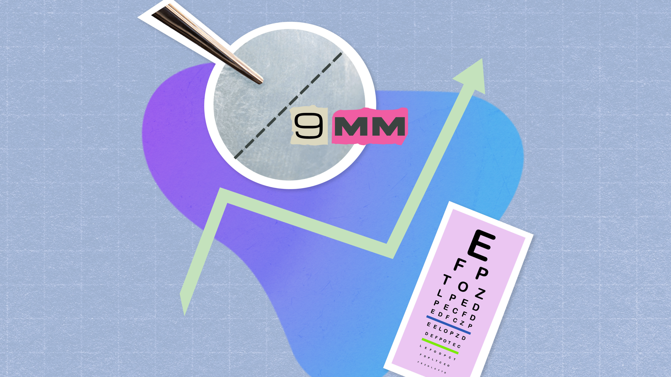
The researchers evaluated whether a double-layered, 9-mm dehydrated AM disc, designed for in-office placement, could safely and effectively promote ocular surface healing and improve vision in patients whose conditions had not responded to standard therapies.
READ MORE: EyeYon Medical’s EndoArt Heads to U.S. Clinical Trial After FDA IDE Approval
Study and design methods
Investigators performed a retrospective chart review of patients treated with a double-layered 9-mm dehydrated AM disc at a tertiary eye care clinic between July 2022 and November 2023.*
Primary outcomes measures focused on changes in uncorrected visual acuity (UCVA) from baseline to the first and last follow-up visits. Secondary outcomes included changes in clinical signs of ocular surface disease and the presence of treatment-related complications.
Ten patients (10 eyes) were included in the analysis. The cohort had a median age of 50 years, and 70% of the patients were male. The mean follow-up duration was approximately 12.9 months, allowing assessment of both short-term and sustained outcomes.*
READ MORE: Ocular Surface Disease, Lipid Layer, and Eyelid Margin Series
Clinical indications and patient outcomes
The amniotic membrane disc was used across several ocular surface indications, including limbal stem cell deficiency in five eyes, persistent epithelial defects in four eyes and aphakic bullous keratopathy in one eye. Despite the diversity of underlying diagnoses, all patients demonstrated clinical improvements following treatment.*
At the first follow-up, the membrane was reported to have successfully integrated in nine of ten eyes (90%). Integration is a key factor in amniotic membrane therapy, as stable adherence allows the membrane to support epithelial healing and mitigate inflammation during recovery.*
Visual outcomes also showed consistent improvement. Mean UCVA improved from 1.69 ± 0.62 logMAR at baseline to 1.22 ± 0.69 logMAR at first follow-up, with further improvement to 0.98 ± 0.70 logMAR at final follow-up. Both changes were statistically significant, with P values of 0.002 and 0.001, respectively.*
READ MORE: Celularity and DefEYE Announce Partnership on Placental-Derived Biologics for Eye Care
Addressing an unmet clinical need
Ocular surface disorders such as limbal cell limbal stem cell deficiency, persistent epithelial defects and aphakic bullous keratopathy are often challenging to treat. These conditions can lead to chronic epithelial breakdown, inflammation, discomfort and progressive visual impairment. While medications like bandage contact lenses and other conventional therapies remain first-line options, patients may continue to experience persistent corneal damage despite treatment.
Amniotic membrane therapy has become an established tool in ocular surface management due to its anti-inflammatory, anti-scarring and regenerative properties. However, traditional amniotic membrane transplantation techniques often rely on thin, single-layer membranes that may degrade quickly and typically require surgical placement using sutures or adhesives in an operating room setting.
This surgical requirement can limit accessibility and increase procedural burden, particularly for patients who require repeat treatments or are poor surgical candidates. The current study explored whether a thicker, double-layered amniotic membrane disc suitable for office-based placement could address these limitations while maintaining clinical effectiveness.
Limitation and future directions
The authors acknowledge limitations related to the retrospective study design and small sample size. Further research involving larger, prospective, controlled studies will be needed to confirm these findings, refine patient selection criteria and compare double-layered amniotic membrane discs with existing single-layer and surgical options.*
The takeaway
While preliminary, the study offers early evidence that a double-layered, dehydrated, 9-mm amniotic membrane disc may provide a safer, effective and less invasive option for managing challenging ocular surface disorders. For clinicians seeking solutions that deliver meaningful healing without sending patients to the operating room, this layered approach may prove to be more than just a thin idea.
Editor’s Note: This content is intended exclusively for healthcare professionals. It is not intended for the general public. Products or therapies discussed may not be registered or approved in all jurisdictions, including Singapore.
*Agarwal M, Huang R, Bareket M, et al. Double layered 9-mm dehydrated amniotic membrane disc in ocular surface disorders: A case series. Eye & Contact Lens. November 14, 2025. ePub ahead of print.